


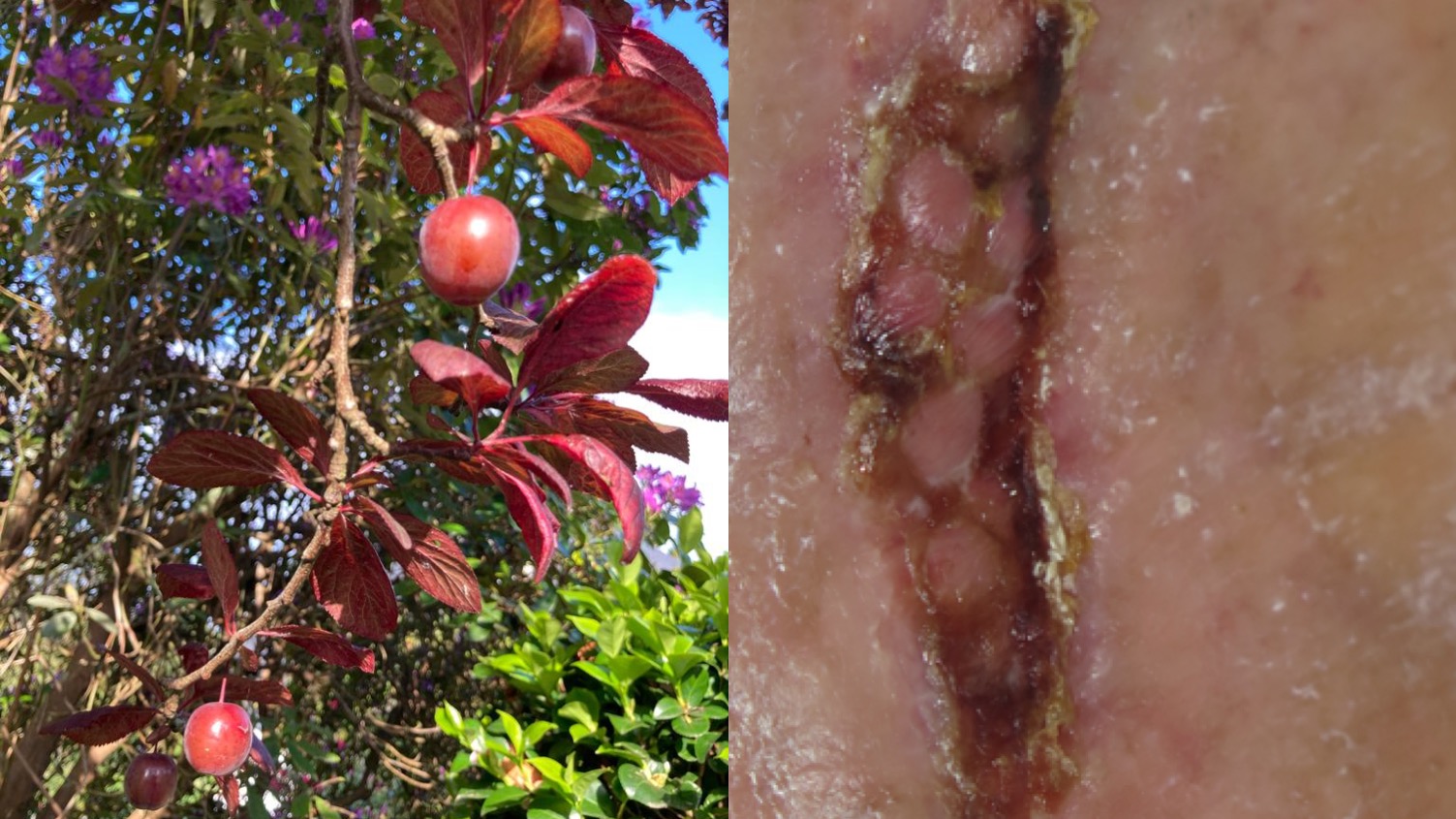
The technique of obtaining and applying grafts in the wound is very simple. However, to be successful with punch grafting, it is essential to know their “color code”;) In this post we are going to understand what the different colors that we can find in dressing changes after the procedure mean.
Graft coloration is associated with neoangiogenesis. As a reminder, neoangiogenesis is the formation of new vessels, which occurs in the first few days after the procedure. Until that time, the grafts do not have their own blood supply, so they must rely on the wound bed for nutrients. If you want to go deeper into the subject, you can read “Braza ME, Fahrenkopf MP. Split-Thickness Skin Grafts. [Updated 2022 May 8]. In: StatPearls [Internet].”
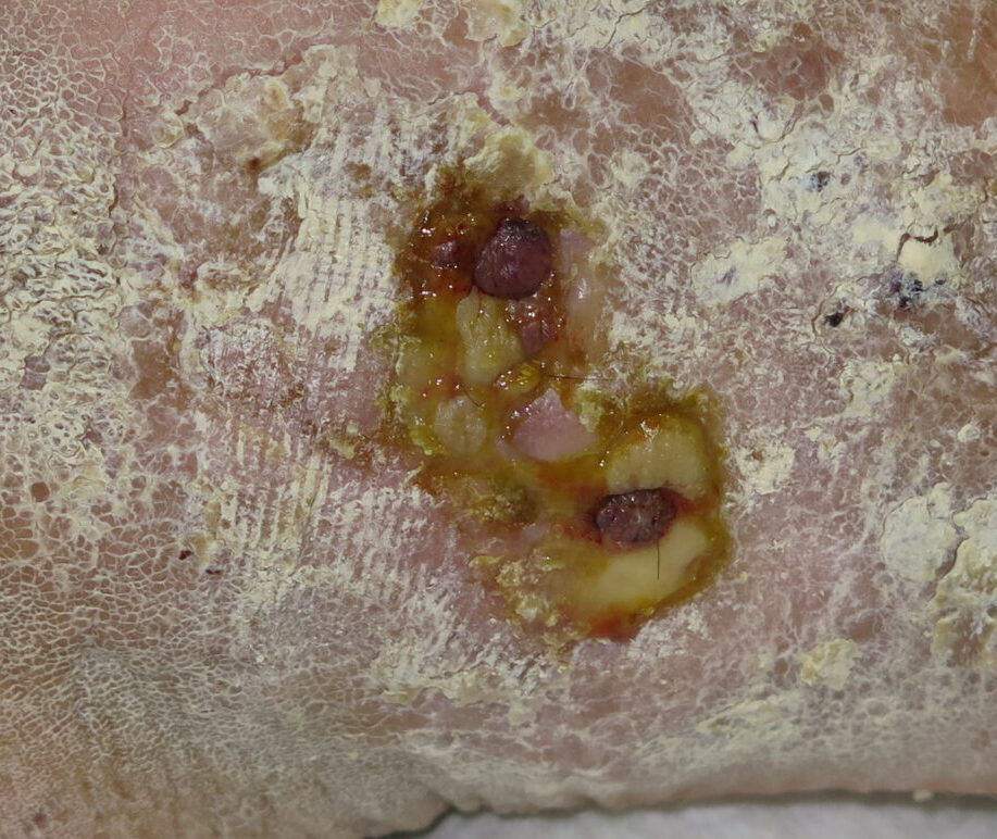
Important notice before continuing: we can find all colors at once in a single wound! (In fact, it is a very frequent occurrence;).
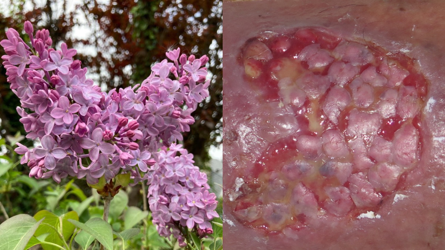
– Yellowish-white:
This pale coloration of the grafts is due to ischemia because a vascular network has not yet been established between the cut vessels on the underside of the graft and the capillary beds in the wound bed. Grafts survive by imbibition, i.e., oxygen and nutrients from the wound bed reach the graft by passive diffusion. This coloration is normal during the first week after the process, usually turning pinkish or bluish in the following days as the graft revascularisation process is completed.

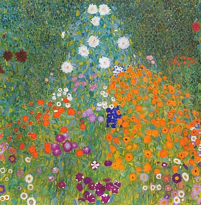
– Blue-purplish:
This is due to venous congestion of the grafts, which is a normal phase in the process of neoangiogenesis and, therefore, a sign of graft attachment. In the following weeks venous drainage of the new vessels occurs and the coloration becomes pinkish.
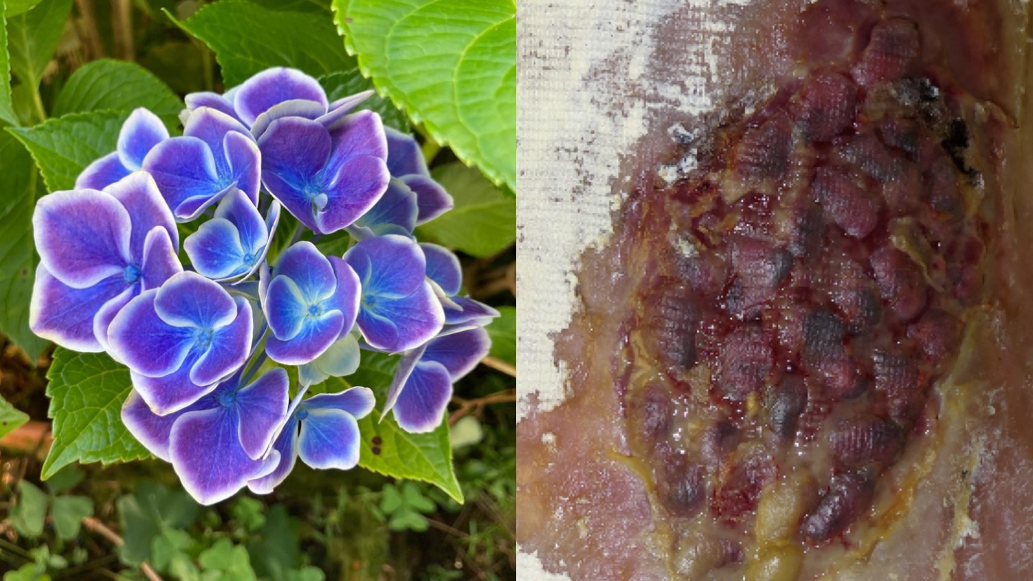
– Pink:
Indicates attachment, i.e. a vascular connection between the graft and the bed. It is common to find pink grafts intermingled with blue-purplish grafts and even pink and violet coloration in the same graft.
– Yellowish-brownish:
This is a sign that the grafts are maintained by imbibition, that is, by diffusion of nutrients from the bed but neoangiogenesis has not occurred. We usually find this coloration when we have grafted a wound in conditions that are not optimal. When we observe it after the first week after the procedure, and the pieces are not fixed in the bed (they are “dancing” in the bed), it indicates that these grafts will not take. Despite this lack of attachment, it is important to keep them in the wound bed, since they release growth factors and cells that promote healing and reduce pain. If we see a stagnation in the healing, from the third week, we can consider the removal of these grafts, which are not attached, and perform a new coverage with punch grafting.
It is also normal to find remains of exudate and scabs between the grafts, which have an epithelialization-promoting function, as well as a protective one. It is therefore essential not to remove this material, which can present different coloration, from yellowish-brownish to garnet-blackish.
After this explanation, you can now understand why the appearance of these grafts in the first dressing change, one week after the procedure, is normal;)
Also available in: Español (Spanish)


